| Jewiki unterstützen. Jewiki, die größte Online-Enzyklopädie zum Judentum.
Helfen Sie Jewiki mit einer kleinen oder auch größeren Spende. Einmalig oder regelmäßig, damit die Zukunft von Jewiki gesichert bleibt ... Vielen Dank für Ihr Engagement! (→ Spendenkonten) |
How to read Jewiki in your desired language · Comment lire Jewiki dans votre langue préférée · Cómo leer Jewiki en su idioma preferido · בשפה הרצויה Jewiki כיצד לקרוא · Как читать Jewiki на предпочитаемом вами языке · كيف تقرأ Jewiki باللغة التي تريدها · Como ler o Jewiki na sua língua preferida |
Datei:Hepatitis B virus v2.png

Originaldatei (843 × 577 Pixel, Dateigröße: 80 KB, MIME-Typ: image/png)
Beschreibung
| Beschreibung |
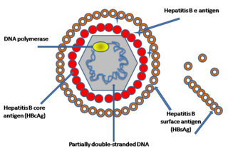
English: Simplified graphical representation of a cross-section of the Hepatitis B virus particle and surface (surplus) antigen, the hepatitis B e antigens (HBcAg) shown are considered not part of the viral particle (quod vide viral nonstructural protein). The structure of the Hepatitis B virus as first described by Dane & al.[1] and Jokelainen, Krohn & al.[2] during 1970. The hepatitis B virion is a complex, double shelled, spherical particle with a 42 nm diameter.[1][2][3]
The virion was initially referred to as the Dane particle.[4] Only after Baruch Blumberg received the Nobel Prize in Medicine during 1976 was it universally accepted that the particle is a virus and the infectious agent of Hepatitis B.
|
| Datum | 14. November 2007 (Original-Hochladedatum) |
| Quelle | Transferred from en.wikipedia |
| Urheber | Created by en:User:GrahamColm. Original uploader was TimVickers at en.wikipedia |
| Genehmigung (Weiternutzung dieser Datei) |
Released into the public domain (by the author). |
| Andere Versionen |
  |

|
Dieses Bild des Typs Biology sollte als Vektorgrafik im SVG-Format neu erstellt werden. Vektorformate haben zahlreiche Vorteile; weitere Information unter Commons:Media for cleanup. Wenn dir eine SVG-Version dieses Bildes vorliegt, so lade diese bitte hoch. Nach dem Hochladen der Datei ist diese Vorlage auf der aktuellen Bildbeschreibungsseite durch die Vorlage {{Vector version available}}, oder kürzer {{Vva}}, zu ersetzen. Es ist empfohlen die neue SVG-Datei „Hepatitis B virus v2.svg“ zu nennen – dann benötigt die Vorlage vector version available (bzw. vva) keinen Parameter.
|
Lizenz
| |
Dieses Werk wurde (oder wird hiermit) durch den Autor, TimVickers auf Wikimedia Commons , in die Gemeinfreiheit übergeben. Dies gilt weltweit. Falls dies rechtlich nicht möglich ist: |
Ursprüngliches Datei-Logbuch
- 2007-11-14 18:14 TimVickers 843×577× (81917 bytes) Simplified drawing of the Hepatitis B virus particle and surface (surplus) antigen
Sources
- ↑ a b c D.S. Dane , C.H. Cameron , Moya Briggs (1970). "Virus-Like Particles in Serum of Patients with Australia-Antigen-Associated Hepatitis". The Lancet 295: 695–698. DOI:10.1016/S0140-6736(70)90926-8.
- ↑ a b c d e f g h i j k l P. T. Jokelainen, Kai Krohn, A. M. Prince and N. D. C. Finlayson (1970). "Electron Microscopic Observations on Virus-Like Particles Associated with SH Antigen". Journal of Virology 6 (5): 685-689. ISSN 1098-5514.
- ↑ a b c d e f The hepatitis B virus. World Health Organisation.
- ↑ a b Almeida J D, Rubenstein D & Scott E J. (1971). "New antigen-antibody system in Australia-antigen-positive hepatitis". The Lancet 298 (7736): 1225–7. DOI:10.1016/S0140-6736(71)90543-5.
- ↑ Bayer, M. E., B. S. Blumberg, and B. Werner (1968). "Particles associated with Australia antigen in the sera of patients with leukemia, Down's syndrome and hepatitis.". Nature (London) 218: 1057-1059.
- ↑ Baruch S. Blumberg, Harvey J. Alter, and Sam Visnich (Jul 1984). "Landmark article Feb 15, 1965: A 'new' antigen in leukemia sera. By Baruch S. Blumberg, Harvey J. Alter, and Sam Visnich". Journal of the American Medical Association 252 (2): 252–7. DOI:10.1001/jama.252.2.252. PMID 6374187. ISSN 0098-7484.
- ↑ Prince, A. M. (1968). "An antigen detected in the blood during the incubation period of serum hepatitis". Proceedings of the National Academy of Science U.S.A. 60: 814-821.
Dateiversionen
Klicke auf einen Zeitpunkt, um diese Version zu laden.
| Version vom | Vorschaubild | Maße | Benutzer | Kommentar | |
|---|---|---|---|---|---|
| aktuell | 20:19, 5. Nov. 2021 | | 843 × 577 (80 KB) | Leonel Sohns | Reverted to version as of 15:17, 8 January 2009 (UTC) New file is erroneous. |
Dateiverwendung
Keine Seiten verwenden diese Datei.